
3D Ultrasound With Color Doppler
What Is 3D Ultrasound With Color Doppler?
There are n-number of infertility causes in women, so diagnosing any probable cause is a complex process. An accurate diagnosis paves the way for the correct approach to treatment. Ultrasounds are one of the classic, reliable, and accurate diagnostic methods for diagnosing infertility causes or routine investigations of pregnancy.
With the advent of technology, we have upgraded our technologies and methodologies to fetch accurate diagnoses from starting fertility treatment to the entire gestation period using color doppler ultrasound in pregnancy. This revolutionary machine gives a clear picture of the fetus and uterus’ internal aspects without incurring any pregnancy risks.
What is 3D Ultrasound, and how is it superior to existing ultrasound methods?
3D ultrasound is a third-generation ultrasound technique that uses high-frequency ultrasound waves and special software to create clear internal images of various organs. The internal images investigate the fetus during pregnancy or uterus lining for fertility treatments.
We follow evidence-based protocols and provide 3D, Color Doppler, and Power Doppler ultrasound investigation that helps gynecologists and fertility experts tailor treatment for each woman seeking treatment.
How does the 3D ultrasound work?
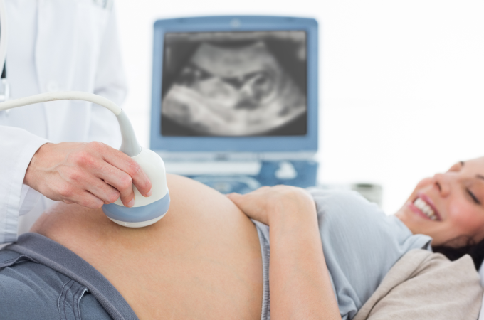
First, the doctor applies a gel on the woman’s tummy and encircles a transducer on the skin. The transducer converts electrical signals into high-frequency sound waves(ultrasound) that strike the internal tissues and organs and reverts to the transducer. The transducer converts the response of ultrasonic waves back to an electrical signal which is simultaneously reflected on the computer screen. The colored doppler superimposes blue, red, and grey shades, which change with internal movements.
The doctor may also do a transvaginal ultrasound to obtain accurate images.
Ultrasound Investigation methods
Transvaginal ultrasound
It is a minimally invasive ultrasound that forms crucial for routine fertility tests. An ultrasound transducer probe is inserted through the vagina to check the uterus clearly. It is helpful in the diagnosis of uterine abnormalities such as cysts, fibroids, inflamed ovaries, etc. It also helps experts count the number of eggs in the ovaries. Ultrasound helps throughout pregnancy with an accurate diagnosis of fetal development.
Pelvic 3D ultrasound
The doctor applies a gel on the abdomen and encircles a transducer probe on the skin. The ultrasound waves fetch corresponding images of the uterus, ovaries, and surrounding tissues on the computer screen.
Hydro-sonography
It is also called fluid sonography. In this, a sterile fluid is injected into the endometrial cavity, and simultaneous transvaginal ultrasounds are taken.
Hyterosalpingo-Contrast- Sonography(HyCoSy)
It is a transvaginal ultrasound recommended for investigating complex infertility in women. It is tolerable, safe, and done for tubular patency or uterine cavity assessment.
Does transvaginal ultrasound impose risk during the last few months of pregnancy?
No, abdominal ultrasound or transvaginal ultrasound (internal ultrasound obtained via the vagina) both are safe and classic investigation methods included as routine testing during the gestation period to estimate fetal development or any other significant changes in the uterus.
It is a painless, radiationless, and cost-effective diagnostic test.
Why Choose Oma Fertility for 3D Ultrasound With Color Doppler?
Ultrasound uses high-frequency sound waves to form corresponding images on the attached screen. It does not only indicate accurate dimensions but also indicates the blood flow rate and direction. This is why a color doppler ultrasound in the 9th month of pregnancy is very helpful for planning deliveries even in emergencies.
OMA IVF values your emotions associated with pregnancy and is always standing by your side in your journey.


